What is the heart?
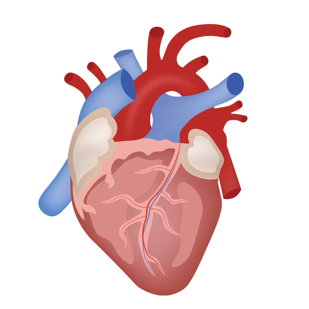
The heart is one of the most important organs in the human body. It is a powerful fist-sized muscle that pumps blood around the body through a network of blood vessels – together, the heart and blood vessels make up the body’s cardiovascular system.
Our hearts beat on average 72 times every minute – over 100,000 times per day. Each minute, vital materials are circulated in our blood and waste products are removed. Each minute counts in helping our body function.
What side is the heart located?
The heart is located in the front of the chest, just behind and slightly left of the breastbone, and between the left and right lungs. Because the heart sits more in the left side of the chest, the left lung is slightly smaller to make room. The ribcage protects the heart.
Heart anatomy
The heart muscle consists of walls, chambers, valves, blood vessels and an electrical conduction system. The entire heart is surrounded by a protective sac called the pericardium that produces fluid to lubricate the heart and protect it from rubbing against other organs.
The heart walls
The heart walls are the muscles that contract and relax to pump blood around the body. There is a layer of muscular tissue (septum) that divides the heart walls into the left and right sides.
The heart walls have three layers: endocardium (inner layer), myocardium (muscular middle layer) and epicardium (protective outer layer that makes up one layer of the pericardium).
The heart chambers
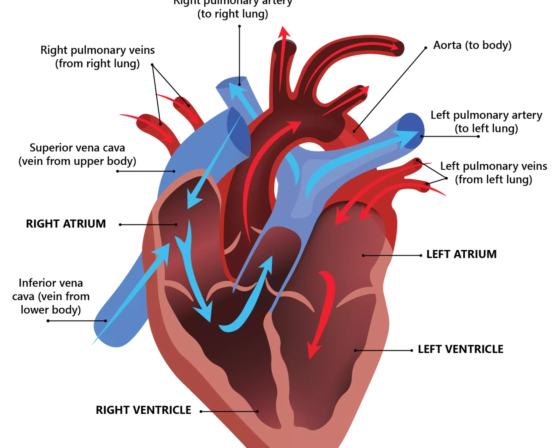
The heart is divided into four chambers: two on the right (right atrium and right ventricle) and two on the left (left atrium and left ventricle). The atria are the upper chambers, and the ventricles are the lower chambers.
- Right atrium: The right atrium receives oxygen-poor blood from the veins and pumps it to the right ventricle.
- Right ventricle: The right ventricle receives the oxygen-poor blood from the right atrium and pumps it to the lungs through the pulmonary arteries, where the blood is loaded with oxygen.
- Left atrium: The left atrium receives the oxygen-rich blood from the lungs through the pulmonary veins and pumps it to the left ventricle.
- Left ventricle: The left ventricle receives the oxygen-rich blood from the left atrium and pumps it to the rest of the body through the aorta. The left ventricle is slightly larger than the right ventricle and is the strongest chamber of the heart. The contractions by the left ventricle are what create blood pressure.
The heart chambers receive oxygen-poor blood from the veins and pump oxygen-rich blood around the body.
Heart valves
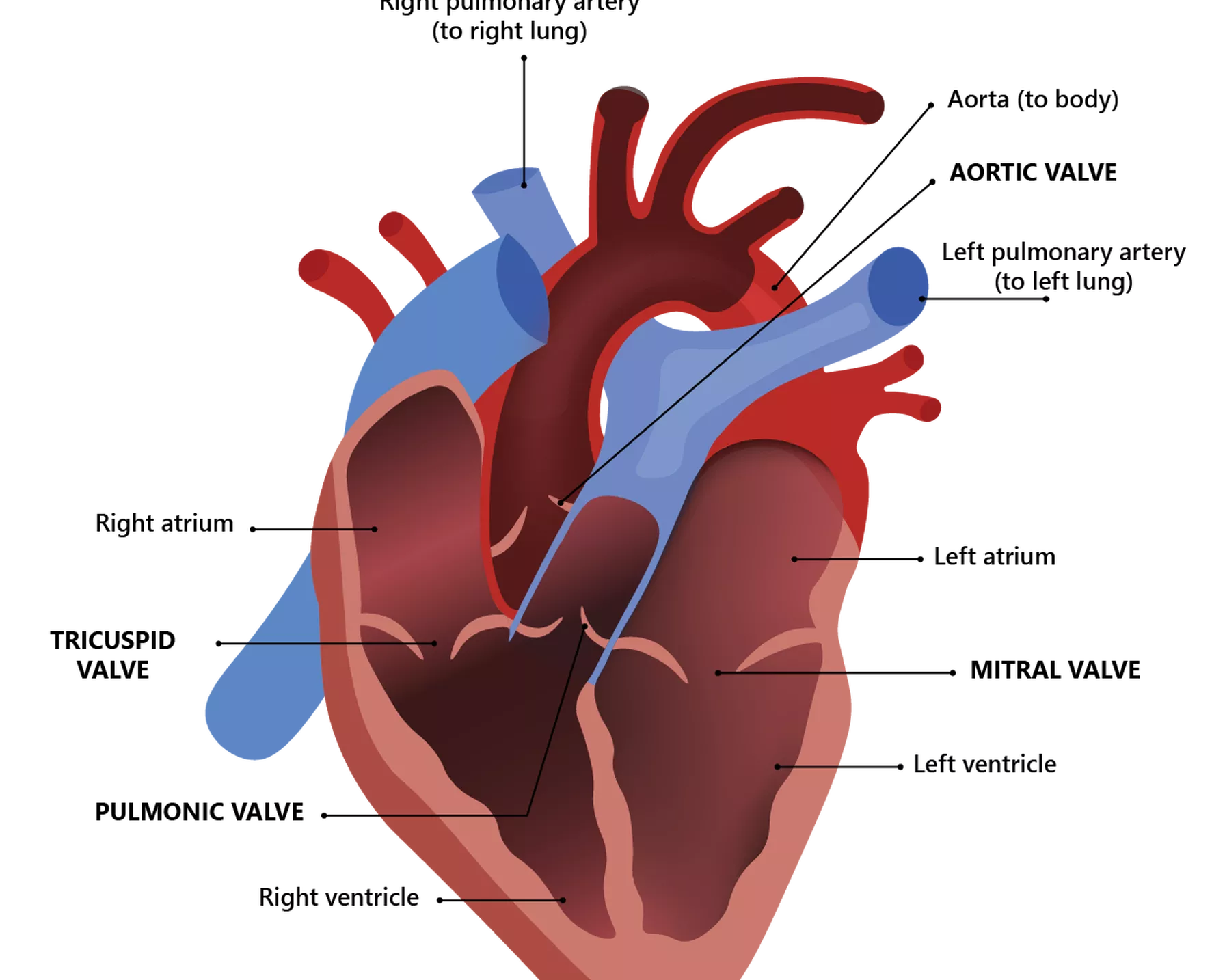
The heart has four valves that act like doors to each of its chambers.
- Tricuspid valve: The tricuspid valve lies between the right atrium and right ventricle.
- Mitral valve: The mitral valve lies between the left atrium and left ventricle.
- Aortic valve: The aortic valve lies between the left ventricle and the aorta, the main artery that carries oxygen-rich blood to the body.
- Pulmonic valve: The pulmonic valve lies between the right ventricle and the pulmonary arteries, the arteries that carry oxygen-poor blood to the lungs to be loaded with oxygen.
The valves open and close to allow blood to flow through the heart, and they keep the blood moving in the correct direction by opening only one way and only when they need to. The valves have flaps that open and close once during each heartbeat.
The heart valves open and close to control the flow of blood through the heart chambers.
Blood vessels
The heart pumps blood through three main types of blood vessels: arteries, veins and capillaries.
- Arteries: Arteries carry oxygen-rich blood from the heart throughout the body. They begin with the aorta, the large artery leaving the heart, and branch several times, becoming smaller and smaller as they carry blood further away from the heart to the organs.
- Veins: Veins carry oxygen-poor blood from the body back to the heart, where it can be pumped to the lungs to be loaded up with oxygen again. Veins become larger and larger as they get closer to the heart.
- Capillaries: Capillaries are the small blood vessels that connect the arteries and veins and through which the body exchanges oxygen-rich blood with oxygen-poor blood. Their thin walls allow oxygen, nutrients and waste products like carbon dioxide to pass to and from the cells of the organs.
The heart requires a supply of oxygen and nutrients to function properly, but it does not receive any nourishment from the blood that it pumps through its chambers. Instead, it receives its own supply of oxygen-rich blood through the coronary arteries, a network of arteries that run along the surface of the heart. In coronary artery disease, fatty plaque builds up in the coronary arteries and prevents the heart from getting the oxygen-rich blood it needs.
Electrical conduction system
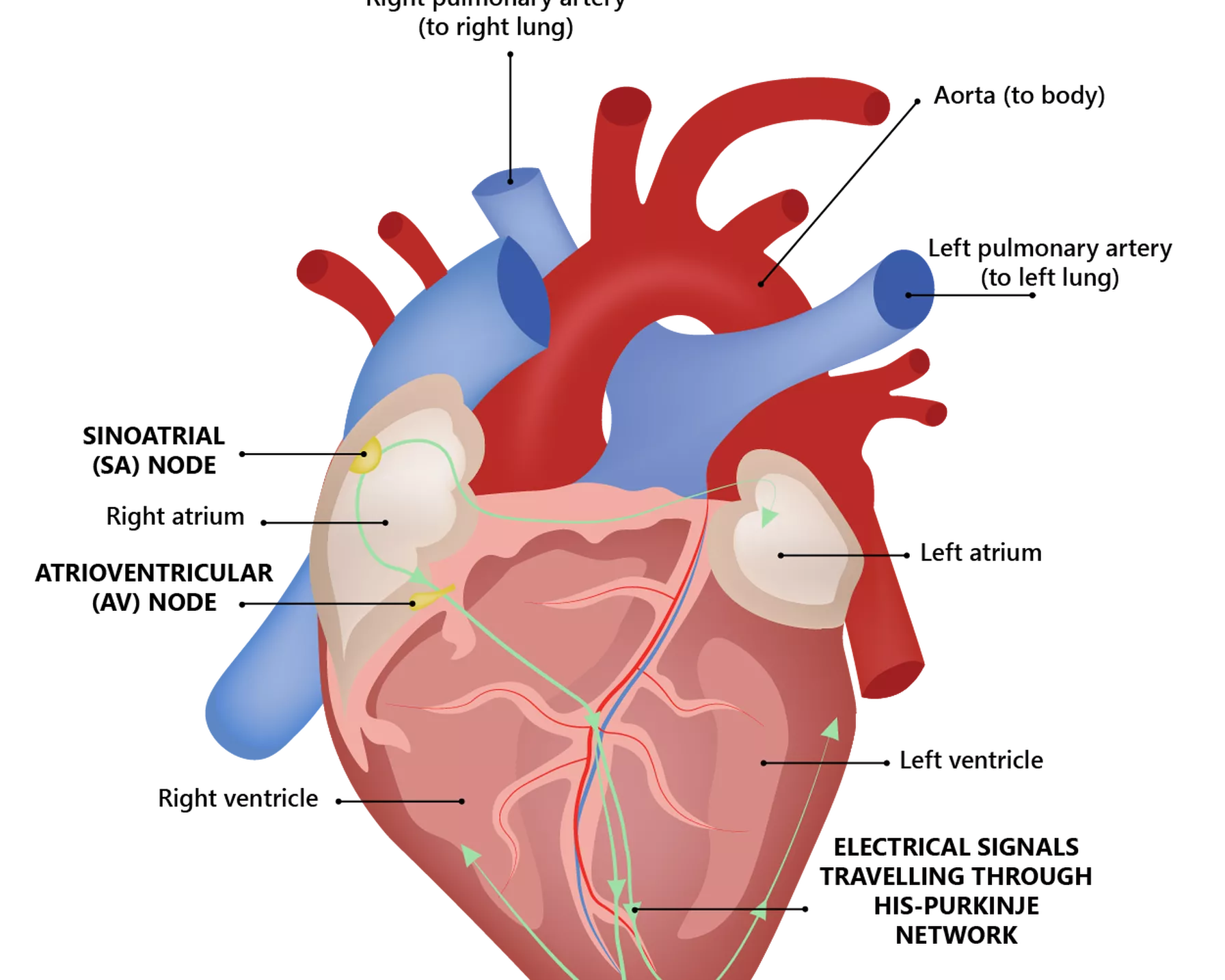
The heart has an electrical conduction system that powers the pumping of the heart, which in turn keeps blood circulating through the body. This system includes two nodes and a specialised network of electrical bundles and fibres.
- Sinoatrial (SA) node: This small bundle of specialised cells located in the right atrium is known as the heart’s natural pacemaker. It sends the signals that make the heart beat and sets the rate and rhythm of the heartbeat.
- Atrioventricular (AV) node: This cluster of cells is located in the centre of the heart, between the atria and ventricles. It carries electrical signals from the heart’s upper chambers to its lower ones.
- His-Purkinje network: This pathway of fibres sends the electrical impulse to the walls of the ventricles, causing them to contract and pump blood out.
The electrical conduction system of the heart triggers its beating.
Heart function
The main function of the heart is to pump oxygen- and nutrient-rich blood around the body. This circulation of blood around the body results in the continuous exchange of oxygen-rich blood with oxygen-poor blood, providing all systems of the body with the oxygen and nutrients they need to function properly.
The heart works with other bodily systems to control heart rate and blood pressure.
How does the heart beat?
Heartbeats are triggered by the electrical impulses that travel through the electrical conduction system of the heart. They occur when the heart contracts and relaxes. The four chambers of the heart work together, alternately contracting and relaxing, to pump blood through the heart. When the ventricles contract, blood is forced into the blood vessels going to the lungs and body. When the ventricles relax, they are filled with blood coming from the atria.
The electrical impulses start in the SA node, located in the right atrium. This electrical activity spreads through the walls of the atria, causing them to contract.
The AV node, located between the atria and ventricles, slows this electrical activity before it enters the ventricles, giving the atria time to contract before the ventricles do.
The electrical activity then passes through the His-Purkinje network to the walls of the ventricles, causing them to contract. This contraction pumps blood out to the lungs and body.
The SA node fires another electrical impulse, and the cycle begins again.
How does the heart work with other organs
The heart works with other systems in the body to control the heart rate, blood pressure and other body functions. The primary systems are the nervous system and endocrine system.
- Nervous system: The nervous system originates in the brain. It controls movements, thoughts and other body systems and processes. The nervous system helps control the heart rate by sending signals that tell the heart when to beat faster or slower, such as during times of stress or rest.
- Endocrine system: The endocrine system is made up of glands that create and release hormones that control nearly all the processes in the body. These hormones tell the blood vessels to constrict or relax, which affects blood pressure. Hormones from the thyroid gland can also tell the heart to beat faster or slower.
Problems with these body systems can affect the heart and lead to cardiovascular disease. For example, diabetes is an endocrine disorder that develops when not enough of the hormone insulin is made, or when insulin in the body does not work as it should. Diabetes is a major risk factor for cardiovascular disease, and it can also cause damage to the nervous system.
Conditions that may affect the human heart
Conditions that affect the heart are among the most common type of disorder. In fact, cardiovascular disease (CVD) is the world’s leading cause of death and disability. CVD refers to all the diseases of the heart and circulation, including the following.
- Atrial fibrillation: Atrial fibrillation is a common, irregular heartbeat condition that is linked to one in three strokes.
- Coronary heart disease: Also known as coronary artery disease, coronary heart disease is a common condition where the major blood vessels to the heart (coronary arteries) become blocked and narrowed, restricting the flow of oxygen-rich blood to the heart.
- Diabetes: Diabetes is a condition in which the body cannot maintain healthy blood glucose levels. People living with diabetes are over twice as likely to develop CVD as the general population.
- Heart attack: A heart attack occurs when the heart is deprived of oxygen due to a blocked artery, and it can lead to death if not treated immediately. It is also known as myocardial infarction.
- Heart failure: Heart failure is when the heart does not work as well as it should in pumping blood and oxygen around the body. Heart failure preserved ejection fraction (HFpEF), a “stiff” type of heart failure where the heart cannot relax properly, is the most common type.
- Stroke: Stroke occurs when the blood supply to the brain is suddenly cut off, such as by a blood clot blocking an artery to the brain. It is a leading cause of disability globally.
The main underlying cause of CVD is atherosclerosis – the build-up of fatty plaques on the walls of the arteries. These plaques are made up of fat, cholesterol, calcium and other substances. Over time, the plaques harden, narrowing the opening of the arteries and restricting blood flow. These atherosclerotic plaques can break, forming a thrombus (blood clot) that can further limit, or even block the flow of blood throughout the body.
How to keep the heart healthy
There are several factors that can impact the health of the heart and increase the risk of heart conditions. Positive lifestyle changes to help keep the heart healthy include the following.
- Eat a nutritious diet that includes a variety of colourful fruit and vegetables, as well as wholegrains and protein.
- Limit intake of processed foods. It’s important to check the nutrition labels of these foods and limit their intake, as they can contain high amounts of saturated fat, trans fat, LDL cholesterol, salt and sugar.
- Avoid soft drinks and other sugary drinks; stay well-hydrated with water instead.
- Choose healthier sources of fat, such as nuts, seeds, avocado and salmon.
- Make regular exercise or physical activity part of your daily routine. Experts recommend at least 30 minutes of moderate-intensity physical activity on most days of the week. Exercise sessions do not need to be done in one block – even small amounts of activity can help.
- Maintain a healthy weight by eating a balanced, nutritious diet and exercising regularly.
- Limit intake of alcohol. Excessive amounts of alcohol can increase the levels of some fats in the blood, reduce the levels of “good” (HDL) cholesterol and increase blood pressure. These can all increase the risk of CVD.
- Quit smoking. Smoking significantly increases the risk of CVD. Both first-hand smoking and long-term exposure to second-hand smoke can damage the arteries that supply blood to the heart and body.
How is HRI protecting the human heart?
HRI conducts groundbreaking research across a broad range of heart-related topics, in our mission to reduce the number of people who die from diseases affecting the heart and to offer a better life for those already suffering from heart disease by developing new treatments and medical devices.
Our Cardiovascular-protective Signalling and Drug Discovery Group is investigating how to repurpose existing drugs for next-generation therapies for heart disease and CVDs.
Our Clinical Research Group is conducting research to detect the earliest signs of heart and blood vessel damage with a view to preventing serious complications later in life due to CVDs such as congenital heart disease.
Our Heart Rhythm and Stroke Prevention Group is investigating strategies to screen for the heart condition atrial fibrillation in the general population, to prevent associated stroke.
